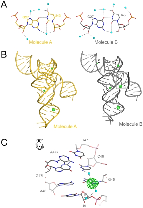
Figure 8. Hydration and metal ion binding of mouse tRNASec.
(A) Hydration of the G27•U43 wobble bps in molecules A (carbon – gold) and B (carbon – silver) of mouse tRNASec, showing a full complement of first-shell water molecules (cyan spheres). Hydrogen bonds are indicated by dashed lines. (B) Anomalous difference Fourier map contoured at the 5 σ level (green mesh) calculated with anomalous differences recorded from a Mn2+-soaked crystal and phases obtained from molecular replacement with the native structure as a search model. Molecule A – gold; molecule B – silver. There are three common Mn2+ sites (1–3) in the two tRNASec molecules. Sites 4 and 5 were found only in chain B. The boxed region is shown in a close-up view in the following panel. (C) Close-up of the boxed region of panel (B). Mn2+ ion (site 4; purple sphere) apparently reinforcing the interaction of U9 (AD-linker) with A48•C45 (first bp of the variable arm).

