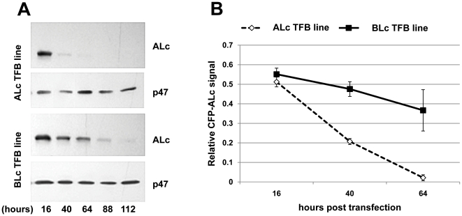
Figure 4. ALc turnover is accelerated in the presence of TFB D5-B8.
(A) ALc expression detected at various times post-transfection in cell lines expressing TFBs. Expression plasmid for CFP-ALc was transfected into cells stably expressing D5-B8 (ALc TFB) or D5-B10 (BLc TFB). Cell lysates were prepared at the indicated time points and resolved by SDS-PAGE. The expression level of CFP-ALc and p47 were monitored by Western blotting using anti-ALc Ab or anti-p47 Ab for detection. (B) Quantitative analyses of ALc expression levels based on scanning densitometry. Western blots such as shown in A were scanned and the signals relative to an internal standard (p47) were calculated and plotted. Data are presented as the average of three sample points ± standard deviation and compared by two-way analysis of variance (ANOVA). The differences between the two cell lines are highly significant (p<0.005) Similar results were obtained in three separate experiments and also using SNAP25 as the internal standard.

