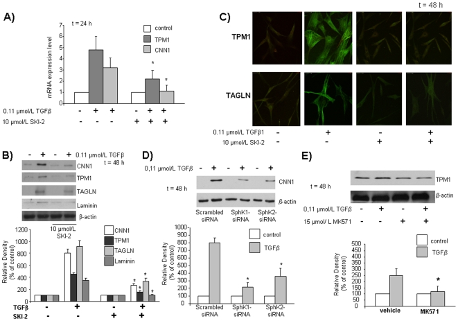
Figure 4. Effect of SphK inhibition (A,B,C), dowregulation (D) and ABCC1 transporter inhibition (E) on the expression levels of SM markers induced by TGFβ treatment in human mesoangioblasts.
Cells were pre-treated with SKI-2 45 min before being incubated with DMEM for 48 h with TGFβ and analyzed by A) Real-Time PCR analysis B) Western blot analysis and C) Confocal microscopy analysis (63× magnification) using anti fluorescein-conjugated specific antibodies. D) Cells were transfected with scrambled-, SphK1-, SphK2-siRNA, stimulated with TGFβ and analyzed by Western blot analysis for CNN1. Lower panel represents densitometric quantification. E) Cells were pre-treated with MK571 45 min in DMEM before being incubated with TGFβ for 48 h and analyzed by Western blot analysis for TPM1. Lower panel represents densitometric quantification.

