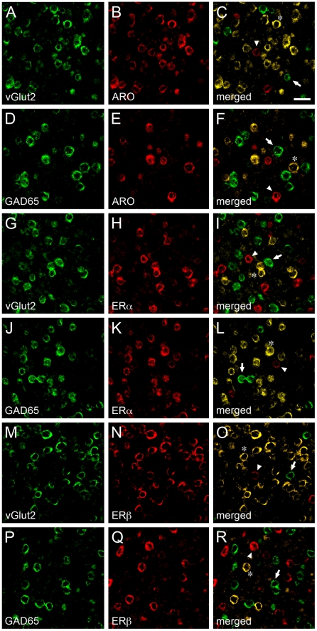
Figure 4. Neurochemical identity and heterogeneity of estrogen-associated circuits in V1.
A–F) Images depicting representative dFISH signal in V1 for vGlut2, a marker for excitatory neurons (A) or GAD65, a marker for inhibitory neurons (D), and ARO (B, E) mRNAs. Note that ARO-positive neurons strongly co-localize with vGlut2 (C), but not GAD65 (F), indicating that estrogen-producing cells in V1 are largely excitatory neurons. G–L) Photomicrographs illustrating dFISH labeling for vGlut2 (G) or GAD65 (J), and ERα (H, K). Notably, whereas few ERα-positive neurons are excitatory (I), the vast majority of these cells co-express GAD65 (L), indicating a GABAergic phenotype. M–R) Images depicting dFISH signal for vGlut2 (M) or GAD65 (P), and ERβ (N, Q) in V1. The merged images (O, R) demonstrate that most ERβ-positive cells are excitatory, but not inhibitory, as revealed by co-localization of vGlut2 (O) and GAD65 (R), respectively. For all merged panels (right-most images in the figure plate), representative double-labeled neurons are highlighted by asterisks. Neurons that are exclusively labeled for either neurochemical cell marker, or markers for estrogen-associated circuits, are depicted by arrows and arrowheads, respectively. Scale bar = 25 µm.

