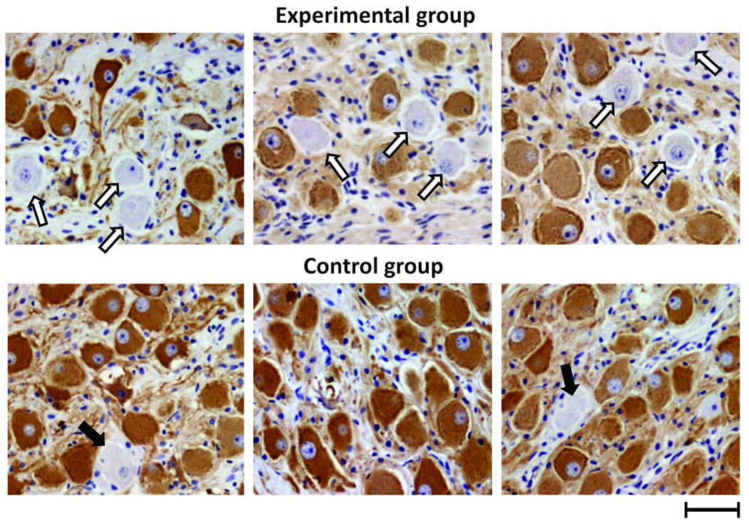
Figure 6.
Tyrosine hydroxylase (TH) immunostaining of left stellate ganglion. Upper panel shows the left stellate ganglion (LSG) of three representative dogs with vagus nerve stimulation (all belong to Group 1). Lower panel shows the LSG of three representative normal control dogs. In experimental dogs, there was a significantly decreased density of TH-positive nerve structures in the LSG and significantly more ganglion cells lacking immunoreactivity to TH (unfilled arrows), which were less common in the control dogs (solid arrows). Scale bar=50 µm.

