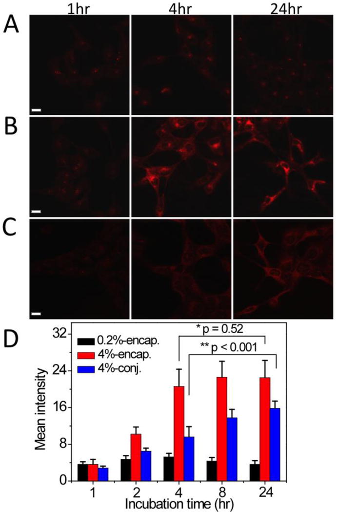
Fig. 5.

Confocal laser scanning microscopy images of H2009 lung cancer cells incubated with PpIX-micelles over time. (A) 0.2% PpIX-encapsulated micelles; (B) 4% PpIX-encapsulated micelles; and (C) 4% PpIX-conjugated micelles. Red fluorescence is from structural moieties containing PpIX. All micelle concentrations were maintained at 100 μg/mL. The scale bars are 20 μm in A-C. (D) The mean fluorescence intensity from 10 cells as a function of incubation time for different PpIX-micelles. The p values were calculated using the Student's t-test and indicated in D between paired groups of comparison.
