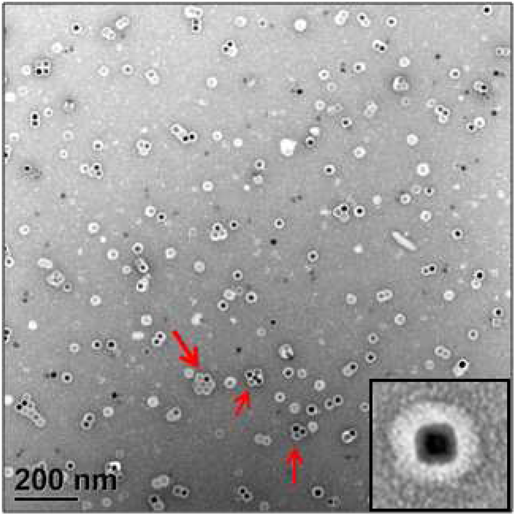
Figure 5.
Negatively stained TEM image of VNPs formed by self-assembling BMV proteins around the 18.6 nm cubic NPs coated with HOOC-PEG-PL. The dark spots are iron oxide NPs. Light rings around NPs are BMV shells including the HOOC-PEG-PL shells. The red arrows illustrate possibly pre-assembled VNP clusters. Inset shows a higher magnification image of a single VNP.

