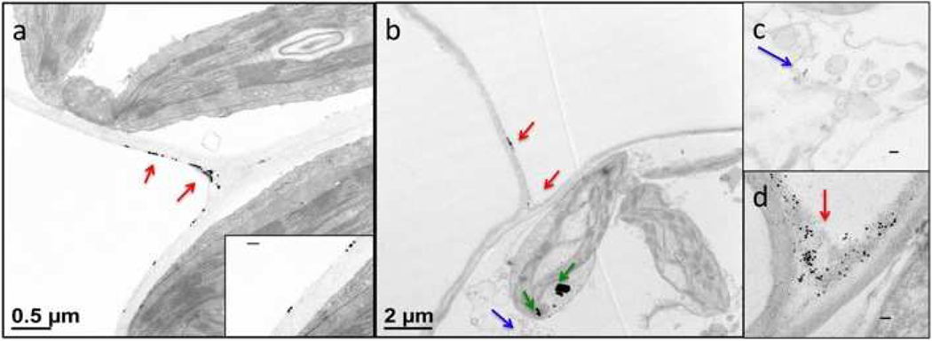
Figure 6.
Stained TEM images of histological preparations of leaf sections. (a) Iron oxide NPs attached to the exterior of the cell wall in the apoplast; red arrows indicate the iron oxide NPs while the enlarged inset shows cubic particles sticking to the cell wall. (b) Magnetic VNPs after entering the plant leaf also gathered at the cell junctions of the apoplast (red arrows) but some particles were present within plant cells (blue arrow). The dark spots inside the chloroplast are indicated by green arrows and are impurities in the staining solution which may have been confused for NPs, were it not for their specific shape. (c) Image of VNPs in the cytoplasm.. (d) Image of VNPs gathered at the cell junction area of the apoplast. All the scale bars in the insets are 100 nm.

