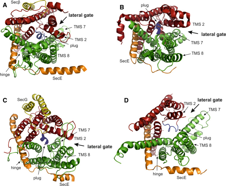
Fig. 1.
Crystal structures of SecYEG in top-view from the cytoplasm. SecY TMS 1–6 (red), TMS 7–10 (green), plug domain (blue), SecE (orange), SecG/β (yellow). a SecYEβ from M. jannaschii (PDB accession code: 1RH5). b SecYE from T. thermophilus co-crystallized with a Fab fragment (not shown) bound to the C5 loop of SecY (2ZJS). c SecYEG from T. maritima co-crystallized with SecA (not shown) (3DIN). d SecYE from P. furiosus. In the crystal, the C-terminal α-helix of a neighboring SecY molecule (not shown) inserts partially into the channel inducing opening of the lateral gate (3MP7)

