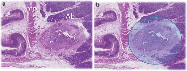
Fig. 2.

Normal histology of the sphincter of Oddi at the portion of the bile duct outside the duodenal wall. a The bile duct is surrounded by the rough and thin sphincter of Oddi, which has partly disappeared on the side of the parenchyma of the pancreas. Ab portion of the bile duct, mp muscle of the duodenum. b Image of the papillary balloon dilation at the portion of the bile duct outside the duodenal wall. A blue and translucent circle, 8 mm in diameter, represents the maximal inflation of the balloon
