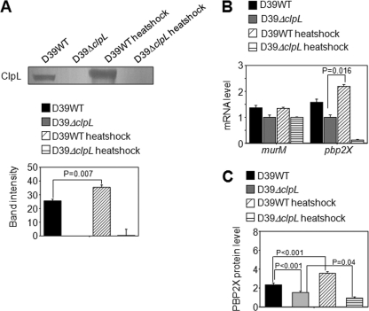
Fig. 6.
Induction of ClpL and PBP2x by heat shock. (A) D39 WT and D39 ΔclpL at the mid-exponential phase (OD550 = 0.3) were heat shocked at 42°C for 30 min. Ten micrograms of bacterial lysate was use to analyze ClpL protein level by Western blot. Colorimetry was used to detect HRP-conjugated secondary antibody used in Western blots. The figure shows representative results of three independent experiments. Band density was analyzed by Photoshop. The figure shows standard deviations from three independent experiments. (B and C) After heat shock, mRNA levels were determined by quantitative RT-PCR (B), and PBP2x protein levels were analyzed by enzyme-linked immunosorbent assay (C) from five (B) and six (C) independent experiments. Significant differences were analyzed by one-way ANOVA.

