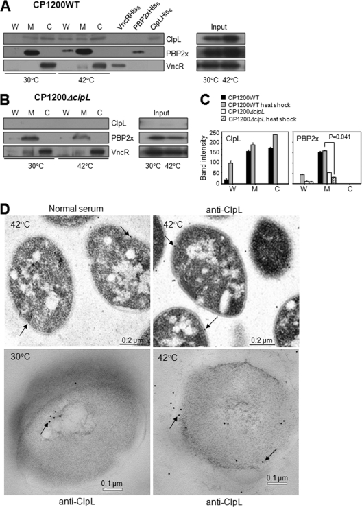
Fig. 8.
Colocalization of ClpL with PBP2x at the cell wall after heat shock. (A to C) After heat shock, the cell wall (W), cell membrane (M), and cytosol (C) of D39 WT (A) and D39 ΔclpL (B) were fractionated. Western blotting using anti-ClpL, anti-PBP2x, and anti-VncR (cytosole marker) antibodies was carried out to localize ClpL and PBP2x. Purified VncR (VncRHis6), PBP2x (PBP2xHis6), and ClpL (ClpLHis6) were added as positive controls. Total cell lysate was used as input. Both chemiluminescence (for ClpL and VncR) and colorimetry (for PBP2x) were used to detect HRP-conjugated secondary antibody used in Western blots. Band density was analyzed by Photoshop. The figure shows the standard deviations from two independent experiments (C). Significant differences were analyzed by ANOVA. (D) Pneumococci cultured at 30°C were heat shocked at 42°C for 30 min. Thin-sectioned samples were treated with preimmune rabbit serum as primary antibody (left and upper panel) or anti-ClpL antibodies, followed by anti-rabbit IgG conjugated to colloidal gold (right, upper and lower panels). The colloidal gold is seen as electron-dense particles (black arrows). The figure shows representative data from three different sections.

