Figure 3.
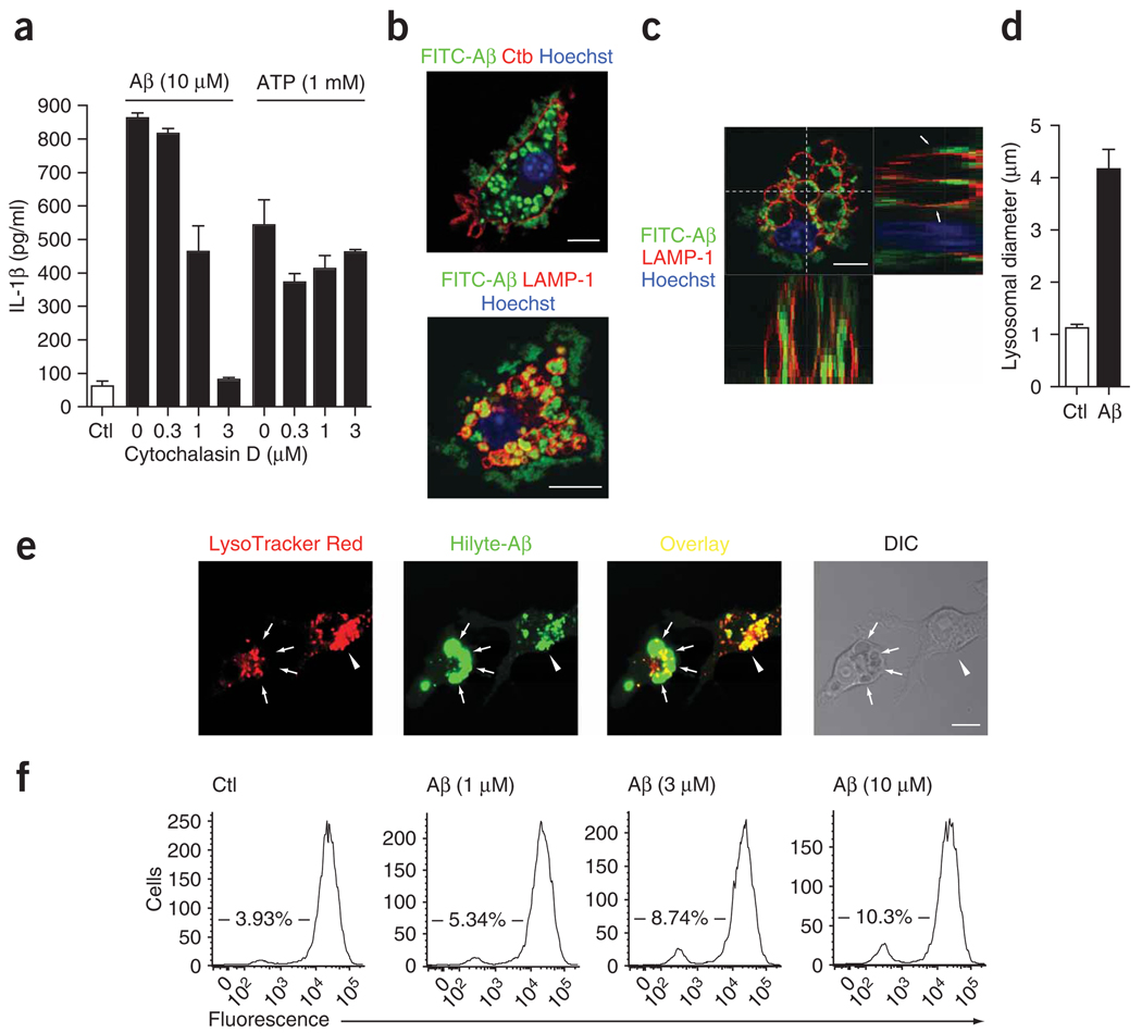
Phagocytosis of Aβ is necessary for IL-1β release and induces lysosomal damage. (a) ELISA of the release of IL-1β into supernatants of wild-type immortalized microglia treated with cytochalasin D during stimulation with Aβ or ATP. (b,c) Confocal microscopy of immortalized microglia incubated for 4 h with FITC-labeled Aβ (10 µM) and then processed for immunocytochemistry; cell membranes were visualized with cholera toxin subunit B and lysosomes were stained with anti-LAMP-1. Arrows indicate LAMP-1-positive lysosomes containing Aβ; z-series (top right and bottom left, c) were taken through the dashed lines (top left, c). Scale bars, 10 µm. (d) Lysosomal diameter in unstimulated cells (Ctl) and cells that incorporated Aβ (n = 50 cells per group). (e) Confocal microscopy of lysosomal integrity in wild-type microglia incubated with acidophilic lysomotropic dye (LysoTracker Red; red), and then stimulated with HiLyte 488–conjugated Aβ (green). Arrowheads indicate phagocytosed Aβ overlaid with the lysomotropic dye in small lysosomes; arrows indicate enlarged Aβ-containing dye-negative lysosomes, indicative of perturbation of lysosomal integrity. Scale bar, 10 µm. (f) Quantification of the loss of fluorescence in e by flow cytometry of wild-type microglia left untreated or treated with Aβ (1, 3 or 10 µM). Numbers in plots indicate percent cells negative for fluorescence. Data are representative of experiments done three times with similar results (error bars (a,d), s.e.m.).

