Figure 6.
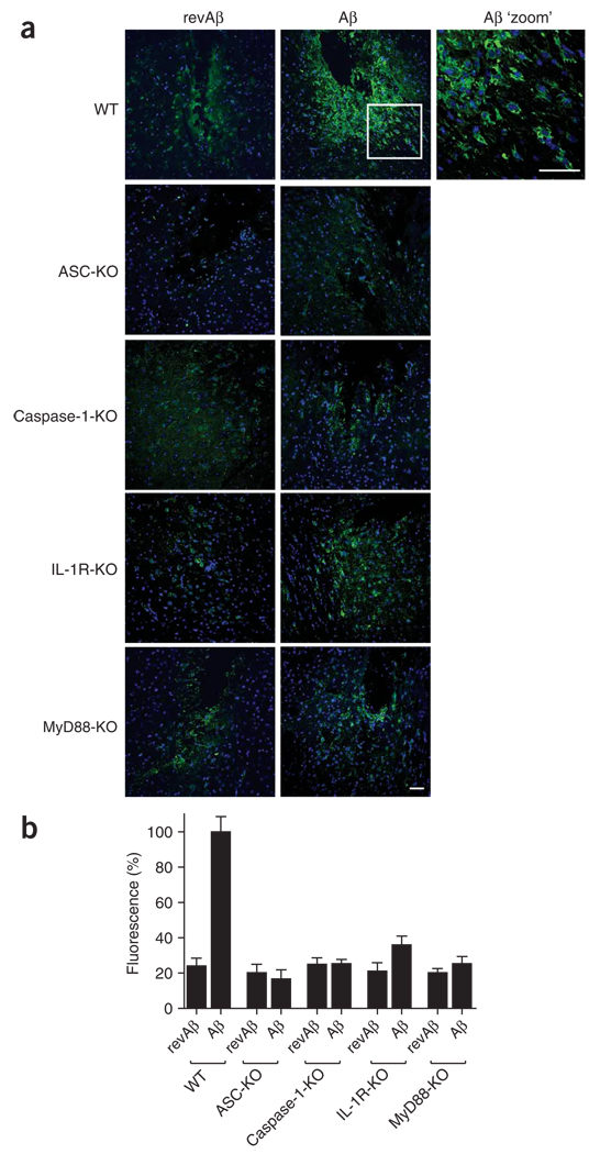
IL-1-mediated pathways contribute to microglial activation induced by Aβ in vivo. (a) Confocal microscopy of coronal brain sections from wild-type mice and mice deficient in ASC, caspase-1, IL-1R or MyD88 at 48 h after stereotactical microinjection of Aβ (1 µg) into the striatum and revAβ into the contralateral hemisphere (control); sections are stained with anti-F4/80 (green) and nuclei are stained with Hoechst 33258 (blue). Scale bars, 50 µm. (b) Quantification of the fluorescence intensity of the F4/80 staining in a to assess recruited microglia and mononuclear phagocytes, normalized to that of wild-type mice treated with Aβ (set as 100%) and presented as the mean and s.e.m. of each group (n = 4 mice per group, with five consecutive sections quantified for each mouse). Data are representative of experiments done twice with each mouse.

