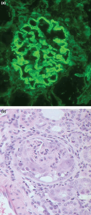
Figure 3.

Kidney tissue at week 12 from CD1 mice with experimental autoimmune glomerulonephritis (EAG) showing (a) strong linear deposits of IgG along the glomerular basement membrane (Direct immunofluorescence; ×300), and (b) marked segmental necrosis of the glomerular tuft with crescent formation (Haematoxylin and Eosin; ×300).
