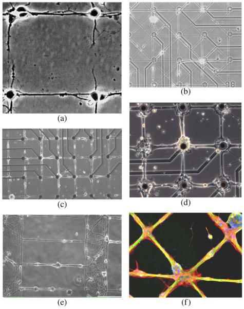
Fig. 2.
Geometric patterns of rat hippocampal neuron growth in culture. (a) Network of individual neurons (laser patterned, Reproduced by permission of Wiley & Sons [14]. (b) Thin line network on 3 μm lines at 14 days in culture. (c) 10 μm wide line network at 55 days in culture. (d) 30 μm lines and 80 μm square nodes at 21 days in culture. (e) Neuropil structure separated by 500 μm with 3 μm wide lines. (f) Cross pattern of 80 μm nodes and 30 μm lines, stained for neurons (green), astroglia (red), and nuclei (blue) (figure was used with permission of the first author for the cover on the Journal of Neural Engineering). In (c), (d) epoxy-silane served as linker for stamped poly-D-lysine and as cytophobic background [31].

