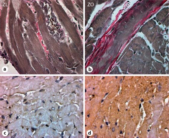
Fig. 2.
The ZO rat heart manifests increased interstitial fibrosis (a, b) compared to the ZL rat heart due to increases in oxidative stress, i.e. 3-nitrotyrosine (c, d). The ZO rat heart (a) displays increased intensity of staining compared to the ZL rat heart (b) with Verhoeff-Van Gieson stain, which stains collagen pink. The ZO rat heart (c) displays increased intensity of immunostaining for 3-nitrotyrosine compared to the ZL rat heart (d).

