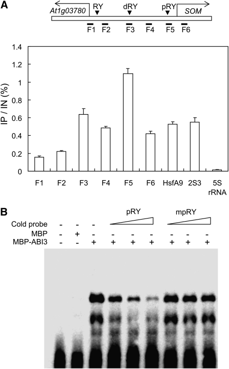
Figure 4.
ABI3 Directly Binds to the SOM Promoter through RY Motifs Both in Vivo and in Vitro.
(A) ChIP analysis showing the direct binding of ABI3 to the SOM promoter in vivo. Top diagram, the SOM promoter and three RY motifs (a proximal RY motif [pRY], a distal RY motif [dRY], and an RY motif near At1g03780 [RY]). Seeds imbibed for 12 h under phyBoff conditions were used for ChIP analysis. F1 to F6 indicate genomic DNA fragments around the SOM promoter tested for enrichment by quantitative PCR (sd, n = 3). IP indicates immunoprecipitated DNA, and IN indicates input DNA.
(B) EMSA analysis showing the direct binding of ABI3 to pRY in the SOM promoter in vitro. MBP-ABI3, ABI3 protein fused to MBP; cold probe, nonlabeled wild-type pRY (pRY) or mutated pRY (mpRY). The increased concentration of competitors (5×, 10×, and 20×) is indicated by triangles.

