
Figure 7.
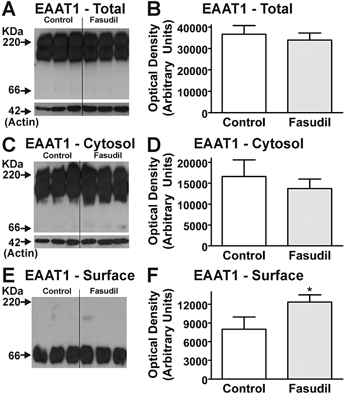
Effects of Fasudil on EAAT1 expression in mouse astrocytes. Western blots demonstrate EAAT1 expression in total cell (A), cytosolic (C) and cell surface (E) fractions of homogenates following treatment with Fasudil (100 µM, 24 h). Labelling of β-actin is included as a loading control. Each lane contains protein extracted from eight culture wells, with different lanes for each treatment being from independent cultures. Quantification of effects of Fasudil on abundance of EAAT1 protein in total cell (B), cytosolic (D) and cell surface (F) fractions of homogenates from cultured astrocytes. *Significantly different from control, P < 0.05, Student's t-test. Data in graphs represent mean ± SEM (n = 3).