Figure 6.

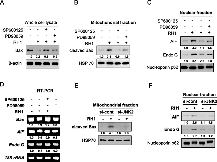
Activation of c-Jun N-terminal kinase (JNK) is required for mitochondrial translocation of cleaved Bax and nuclear translocation of apoptosis-inducing factor (AIF) and endonuclease G (Endo G) in response to 2,5-diaziridinyl-3-(hydroxymethyl)-6-methyl-1,4-benzoquinone (RH1) treatment. (A) NAD(P)H quinone oxidoreductase 1 (NQO1)+-MDA-MB-231 cells were treated with 10 µM RH1 for 9 h in the presence or absence of 30 µM SP600125 or PD98059. Whole cell lysates were subjected to Western blot analysis using anti-Bax and -β-actin antibodies. The relative density value of each band is shown below the Western blot. The data are from a typical experiment that was conducted three times with similar results (mean values, P < 0.05). (B) NQO1+-MDA-MB-231 cells were treated with 10 µM RH1 for 9 h in the presence or absence of 30 µM SP600125 or PD98059. Mitochondrial fractions were prepared and subjected to Western blot analysis using anti-Bax and -mitochondrial HSP70 antibodies. The relative density value of each band is shown below the Western blot. The data are from a typical experiment that was conducted three times with similar results (mean values, P < 0.05). (C) NQO1+-MDA-MB-231 cells were treated with 10 µM RH1 for 9 h in the presence or absence of 30 µM SP600125 or PD98059. Nuclear fractions were prepared and subjected to Western blot analysis using anti-AIF, -Endo G and -Nucleoporin p62 antibodies. The relative density value of each band is shown below the Western blot. The data are from a typical experiment that was conducted three times with similar results (mean values, P < 0.05). (D) NQO1+-MDA-MB-231 cells were treated with 10 µM RH1 for 9 h in the presence or absence of 30 µM SP600125 or PD98059, and RT-PCR products were obtained using specific primers recognizing Bax, AIF and Endo G mRNA, and 18S rRNA. The relative density value of each band is shown below the RT-PCR data. The data are from a typical experiment that was conducted three times with similar results (mean values, P < 0.05). (E) NQO1+-MDA-MB-231 cells were treated with 10 µM RH1 for 9 h in the presence or absence of siRNA targeting JNK2. Mitochondrial fractions were prepared and subjected to Western blot analysis using antibodies against Bax and mitochondrial HSP70. The relative density value of each band is shown below the Western blot. The data are from a typical experiment that was conducted three times with similar results (mean values, P < 0.05). (F) NQO1+-MDA-MB-231 cells were treated with 10 µM RH1 for 9 h in the presence or absence of siRNA targeting JNK2. Nuclear fractions were prepared and subjected to Western blotting using antibodies against AIF, Endo G and Nucleoporin p62. The relative density value of each band is shown below the Western blot. The data are from a typical experiment that was conducted three times with similar results (mean values, P < 0.05).
