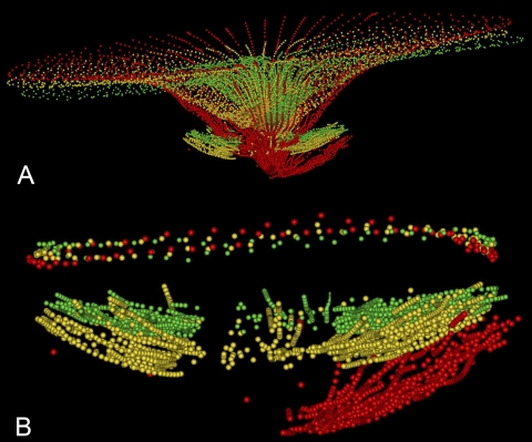
Figure 10.
ILM and ALCS displacement in an EG eye (18389 in Fig. 9). (A) ILM and ALCS delineations are shown, with BL1 (green), FU1 (yellow) and FU2 (red). (B) The ALCS delineations are magnified, anchored at the level of the NCO (shown above the ALCS delineations).

