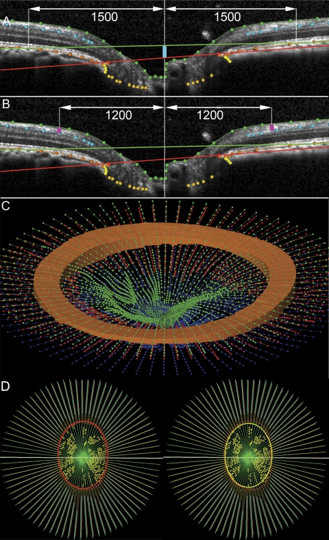
Figure 3.
Parameterization of spectral domain optical coherence tomography volumes. (A) A horizontal B-scan. The primary reference plane (red line) is located at the level of the NCO. A secondary, peripheral reference plane is located at Bruch's membrane at 1500 μm eccentricity from the NCO centroid. The mean depth of the NCO is measured from the secondary reference plane to the NCO (turquoise line). (B) Mean RNFLT is measured between the ILM and the posterior surface of the RNFL at 1200-μm eccentricity from the NCO centroid (pink lines). (C) Three dimensional view to illustrate the RNFLV, shown as an orange circumpapillary cylinder bound by the internal limiting membrane (green) between 1200 and 1500 μm eccentricity from the NCO centroid and the posterior surface of the RNFL (in this panel shown here in red and in A and B in turquoise). (D) The en face view of a fully delineated volume is shown. Left: the red ellipse defines the NCO area. Right: BT area is bound by the yellow ellipse, located at the innermost BT delineations.

