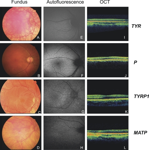
Figure 2.
Fundus photography, FAF, and OCT analysis in four patients with mutations in different OCA genes: P3 (A, E, I), carrying TYR mutation; P36 (B, F, J) bearing a P mutation; P40 (C, G, K) with a TYRP1 mutation; and P41 (D, H, L) showing an MATP mutation (see Table 1). (A–D) Fundus photographs: choroidal vessels are visible in the macula in P3 (A) and P41 (D), but they are not visible in P36 (B) and P40 (C); (E–H) FAF: please note the absence of macular pigment in P3 (E) and P41 (H) and the presence of macular pigment in P36 (F) and P40 (G). (I–L) OCT of the posterior pole crossing the fovea, showing absence of the foveal pit in P3 (I) and P41 (L) and presence of the foveal pit in patients P36 (J) and P40 (K).

