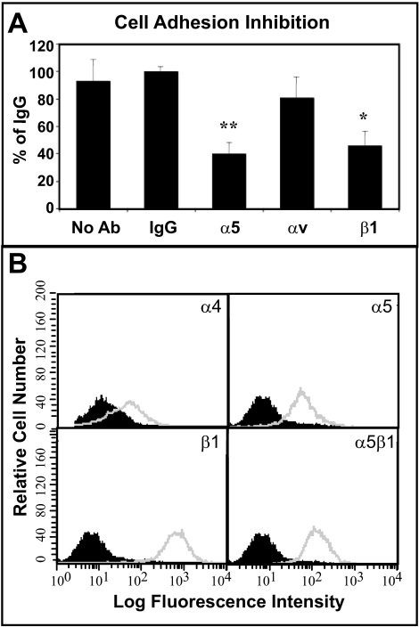
Figure 2.
HTM cell adhesion via α5β1 integrin and integrin expression. (A) The α5 and β1 integrin blocking antibodies significantly inhibited cell attachment to the CBD of fibronectin (*P = 0.0004, **P < 0.0001 compared to IgG control). The αv integrin blocking antibody had no significant effect. Differentiated A7–1 cells were plated on the CBD for 1 hour in the absence or presence of integrin blocking antibodies or control IgG. Bound cells were colorimetrically quantified using toluidine blue. Data are the mean percentage of IgG ± SEM. (B) FACS analysis of integrin levels in differentiated A7–1 cells. Cells were labeled with antibodies against various integrin subunits. Non-immune IgG controls are shown as black peaks, integrin expression is shown in gray.

