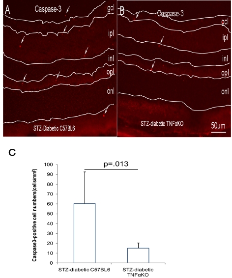
Figure 5.
Reduction of activated caspase-3–positive cells in STZ-induced diabetic TNFα (KO) mice. Mice with diabetes of 3 month's duration were used. The apoptotic cells were indicated by activated caspase-3 staining in STZ-induced diabetic TNFα+/+ (A) or TNFα−/− (B) mice. (C) The quantification of activated caspase-3–positive cells. The results are expressed as the mean ± SE of five independent experiments. Arrows: apoptotic cells. gcl, ganglion cell layer; ipl, inner plexiform layer; inl, inner nuclear layer; opl, outer plexiform layer; onl, outer nuclear layer.

