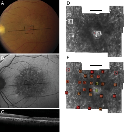
Figure 3.
Fundus photograph (A) and AF (B) of patient 3 (Gly208Asp) shows RPE mottling diffusely throughout the macula. (A, B, black lines) Region imaged with AOSLO and SD-OCT, respectively. (C) SD-OCT through fixation reveals outer nuclear, inner segment, and outer segment layer loss eccentric to a central region of relatively preserved outer retinal structure, where highly reflective features are present in the outer segment/RPE junction layer (arrow). (D) High-resolution images of macular cone structure show cone spacing is irregular and normal (0.95 SD above the mean) in regions but increased by >5 SD above the mean in regions near the fovea. Red dot: fixation. Fundus-guided microperimetry shows >2 log units sensitivity loss (indicated by 0) in regions of heterogeneous AF, with 1 to 2 log units sensitivity loss within 1° of fixation (E). Hyperreflective cones are present at fixation (see Fig. 6C, left). Scale bar, 1°.

