Figure 2.
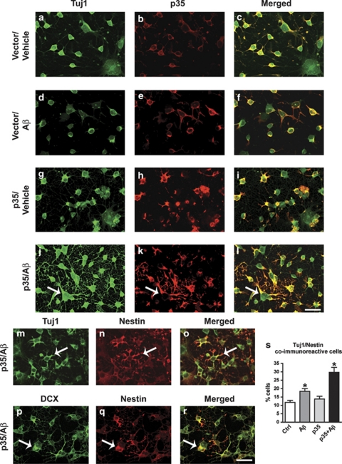
Morphological and maturation alterations in NPC-derived neural progeny expressing p35 and treated with Aβ. Differentiating NPCs were infected with an adenoviral vector expressing p35 or vector control on day 2 of differentiation, and then treated with 1 μM Aβ or vehicle control on day 3 for 24 h, followed by fixation with paraformaldehyde. Samples were processed for double-immunolabeling with antibodies against p35 and markers of proliferative and neuronal differentiation status (neuronal, β-III tubulin (Tuj1), and doublecortin (DCX); progenitor cells, nestin). (a–f) Double-labeling analysis with Tuj1 and p35 antibodies in vector-infected NPC-derived neural progeny treated with vehicle control (a–c) or Aβ (d–f) for 24 h. (g–l) Double-labeling analysis with Tuj1 and p35 antibodies in p35-expressing NPC-derived neural progeny treated with vehicle control (g–i) or Aβ (j–l) for 24 h. (j–r) In combined p35/Aβ-treated cultures, progeny acquired an abnormal stellate morphology and cells were co-immunoreactive for markers of proliferation stages (nestin) and neuronal (Tuj1 and DCX) lineage (arrows). (s) Quantitative analysis of proportion of NPC-derived neural progeny co-labeled with Tuj1 and nestin antibodies. Scale bar=15 μm. *P<0.05 compared with vehicle-treated controls by one-way ANOVA with post hoc Dunnett's test (N=3)

