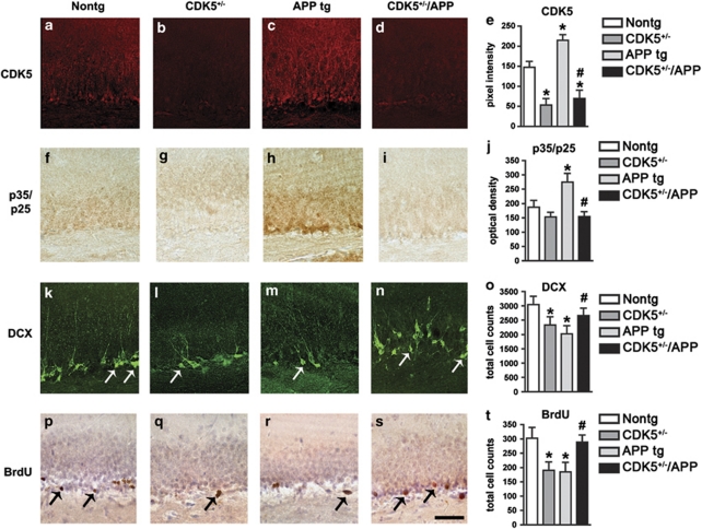
Figure 5.
In vivo genetic modulation of CDK5 rescues neurogenic deficits in the hippocampus of APP tg mice. Nine-month-old wild-type control mice (non-tg), CDK5 heterozygous deficient (CDK5+/−), APP tg mice, or crosses (CDK5+/−/APP) were treated with BrdU. Brain sections were processed for immunohistochemical analysis with antibodies against CDK5, p35/p25, doublecortin (DCX), or BrdU. All images are from the dentate gyrus of the hippocampus. (a–e) Immunohistochemical analysis of the levels of CDK5 immunoreactivity in wild-type, CDK5+/−, APP tg mice, or CDK5+/−/APP crosses. (f–j) Immunohistochemical analysis of the levels of p35/p25 immunoreactivity in wild-type, CDK5+/−, APP tg mice, or CDK5+/−/APP crosses. (k–o) Immunohistochemical analysis of the numbers of DCX-immunoreactive cells (arrows) in wild-type, CDK5+/−, APP tg mice, or CDK5+/−/APP crosses. (p–t) Immunohistochemical analysis of the numbers of BrdU-immunoreactive cells (arrows) in wild-type, CDK5+/−, APP tg mice, or CDK5+/−/APP crosses. Scale bar=50 μm. *P<0.05 compared with wild-type controls by one-way ANOVA with post hoc Dunnett's test. #P<0.05 compared with APP tg mice by one-way ANOVA with post hoc Tukey–Kramer test. N=4 animals per group

