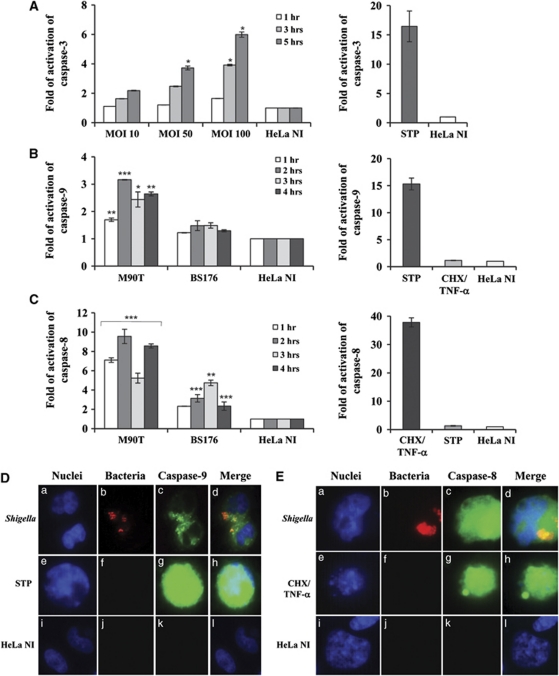
Figure 2.
Caspase activation in HeLa cells infected with Shigella. (A) Kinetics of caspase-3 activation following infection of HeLa cells with M90T at different MOIs as above. (B) Caspase-9 and (C) caspase-8 activity in HeLa cells infected with M90T and its noninvasive variant BS176 (MOI of 100) at the reported time points. HeLa cells treated for 4 h with STP (2 μM) or with CHX (10 μg/ml) plus TNF-α (100 ng/ml) for 12 h were used as a control of caspase-9/3 and caspase-8 activation, respectively. HeLa NI, non-infected HeLa cells. Report assay data correspond to the mean±S.D. (triplicate determinations) and are representative of three independent luminometric assays. *P<0.05, **P<0.01, ***P<0.001 after Student's t-test. (D and E) Immunofluorescence analysis of caspase-9 (D) and caspase-8 (E) maturation in HeLa cells infected with M90T-DsRed (MOI 100) at 2 h of incubation p.i. (a, b, c, d panels) Treated with STP or CHX plus TNF-α, as above (e, f, g, h panels in D and E, respectively) and uninfected (i, j, k, l panels). Cells were processed for labeling with fluorescent-coupled LEHD inhibitor to the activated form of caspase-9 (FLICA caspase-9 (FAM-LEHD-FMK)) or fluorescent-coupled LETD inhibitor to the activated form of caspase-8 (FLICA caspase-8 (FAM-LETD-FMK)) and DAPI. Shigellae, expressing DsRed plasmid vector, are stained in red. Magnification: × 40

