
Figure 1.
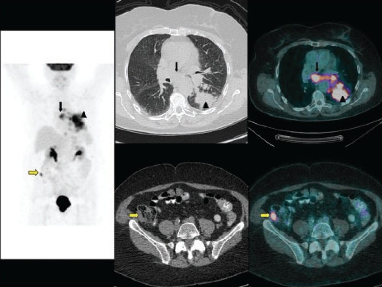
A 73-year-old woman who came for initial staging of non-small cell lung cancer. Maximum-intensity-projection (MIP) image (left panel), CT images (middle panels) and fused images (right panels) of 18F-FDG PET/CT show the primary tumor (arrow head) with mediastinal nodal metastases (black arrow). Incidental right iliac fossa small focal uptake (yellow arrow) is noted, which cross-correlated to a small soft tissue lesion in the cecum and turned out to be a synchronous primary adenocarcinoma.