
Figure 3.
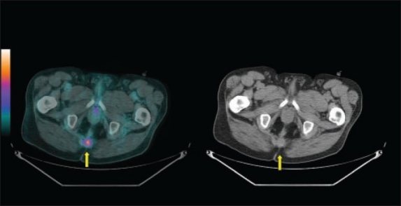
A 72-year-old man who had unexplained elevation of CEA during his follow-up after treatment of anorectal cancer. Fused (left panel) and CT (right panel) images of 18F-FDG PET/CT scan showed recurrent avid disease at the residual surgical bed soft tissue density (yellow arrow).