
Figure 5.
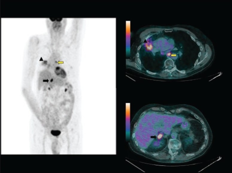
A 68-year-old man who came for initial staging of non-small cell lung cancer. MIP image (left panel) and fused images (right panel) of 18F-FDG PET/CT showed the primary tumor (arrow head) with mediastinal nodal involvement (yellow arrow) and extra-thoracic right adrenal metastasis (black arrow).