
Figure 6.
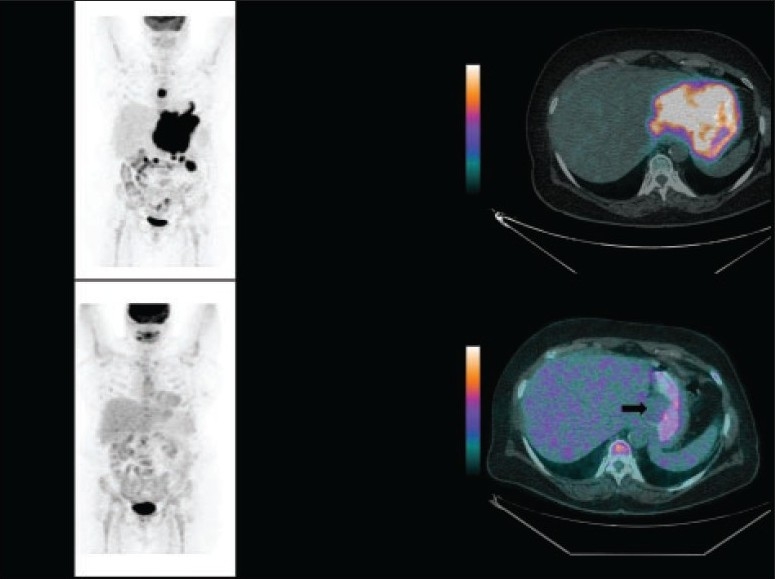
A 66-year-old woman diagnosed with Hodgkin lymphoma. The 18F-FDG PET/CT study (left and right upper panels) for initial staging showed nodal involvement above and below the diaphragm. 18F-FDG PET/CT after four cycles of chemotherapy (left and right lower panels) showed complete metabolic resolution of the disease with small non–FDG-avid residual soft tissue (black arrow on the fused image).