
Figure 7.
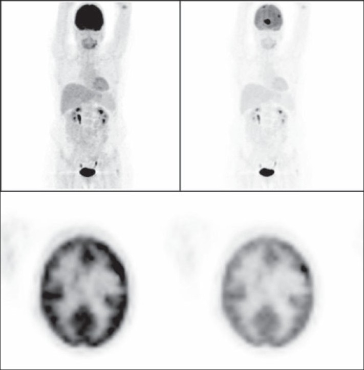
A 62-year-old woman with history of breast cancer. The left column images (MIP and axial PET images) show the normal-intensity images, which could hide metastatic deposits and give a false-negative result due to the physiological high background intensity of the brain. The same images after reducing their intensity on the right column show the metastatic deposits.