Figure 5.
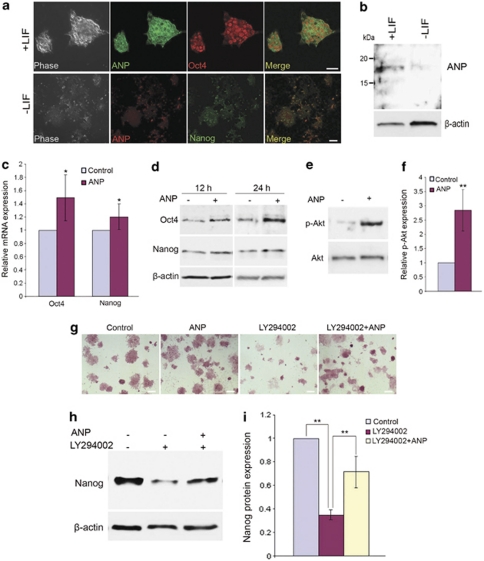
ANP is expressed in murine ES cells, and exogenous ANP stimulates ES cell pluripotency factors. (a) Upper panels, double-immunofluorescence images of ES cells cultured in the presence of LIF, stained with antibodies against the ES cell markers, Oct4 and ANP. Lower panels, double-immunofluorescence images of ES cells cultured in the absence of LIF for 5 days, stained with antibodies against Nanog and ANP. It must be noted that ANP signals are downregulated upon differentiation. (b) Western blot analysis for ANP of ES cells treated as described in panels a and b. (c) Real-time PCR analysis of mRNA levels for pluripotency genes 12 h after treatment of ES cells with 2.5 μ ANP. (d) Western blot analysis of Oct4 and Nanog in ES cells treated with ANP for 12 or 24 h. β-Actin is shown as a control for loading. (e) Western blot analysis for phosphorylated Akt (Ser473; p-Akt) and total Akt 12 h after treatment of ES cells with ANP. (f) Relative levels of the p-Akt protein were quantified after normalized to total Akt. (g) ES cells were treated with LY294002, ANP or ANP+LY294002 for 3 days, followed by staining for alkaline phosphatase (AP). (h) Western blot analysis of Nanog in ES cells treated with LY294002 or ANP+LY294002 for 18 h. (i) Quantitative analysis of the western blots as shown in panel h. Data represent mean±S.D. (n=3). *P<0.05 or **P<0.01 (two-tailed t-test)

