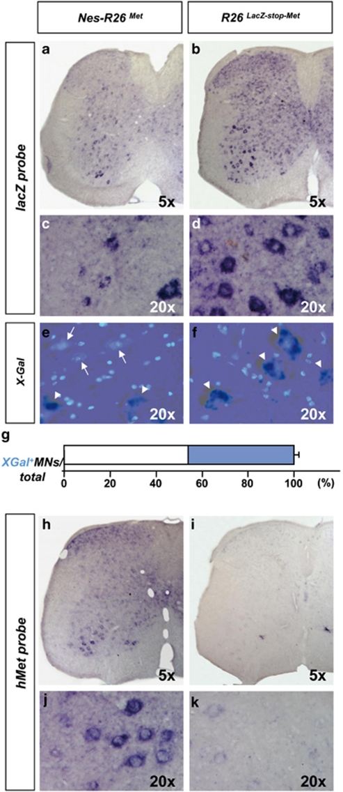
Figure 3.
Molecular analysis of Nes-R26Met and R26LacZ−stop−Met mice in adult lumbar spinal cords on transversal sections. (a and b) In situ hybridization using lacZ riboprobes. (c and d) Enlarged view of the ventral regions, showing lacZ expression in distinct cell types characterized by large cell nuclei resembling MNs in R26LacZ−stop−Met and a decrease in its levels after nestin-cre-mediated recombination in Nes-R26Met. (e and f) Section showing the overlap of X-Gal activity and DAPI staining in lumbar spinal cords. X-Gal-negative cells with large nuclei were predominantly found in Nes-R26Met, but not in R26LacZ−stop−Met transgenics. Arrows and arrowheads points to X-Gal-negative and X-Gal-positive cells with large nuclei, respectively. (g) Quantification analysis of X-Gal-positive (blue) and X-Gal-negative cells with large nuclei (resembling MNs; white) in Nes-R26Met mice, showing that nestin-cre-mediated lacZ excision occurred in approximately 56% of these cells. Values are expressed as means±S.E.M. (h–k) Expression of the met chimeric transgene in lumbar spinal cords. Panels (j and k) show an enlarged view of ventral spinal cords and met transgenic expression in cells resembling MNs

