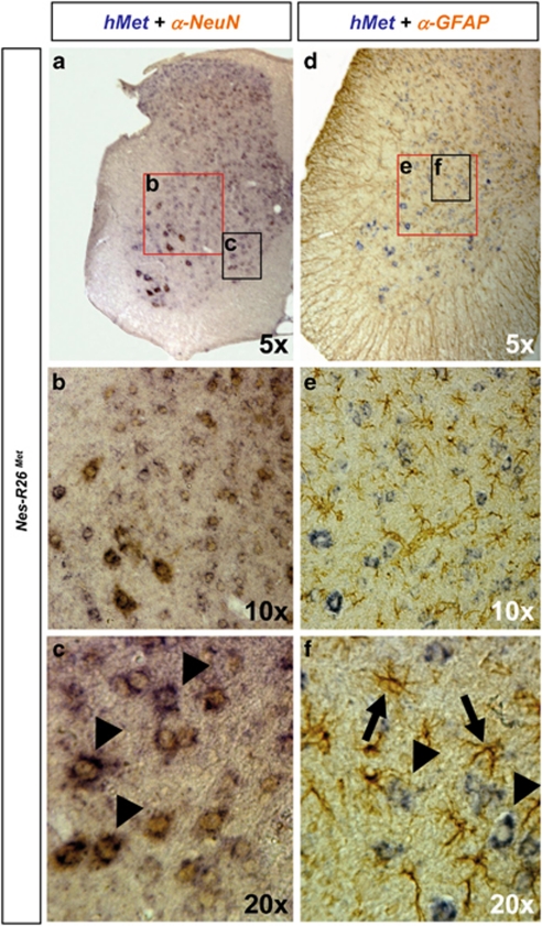
Figure 5.
Subcellular localization of chimeric met transcripts in lumbar spinal cords of adult Nes-R26Met mice. (a–c) Colocalization studies of exogenous met transcript with NeuN protein showing met expression in neuronal cell types. (d–f) Colocalization studies of exogenous met transcript with GFAP protein showing that transgenic met is not predominantly expressed in astrocytes. Panels (b and e), panels (c and f) correspond to an enlarged view of spinal cord areas indicated by red and black rectangles in (a and b), respectively. Arrowheads in (c) point to MNs co-expressing chimeric met transcript and NeuN protein. Arrows and arrowheads in (f) indicate GFAP-positive astrocytes and MN expressing chimeric met transcripts, respectively

