Figure 7.
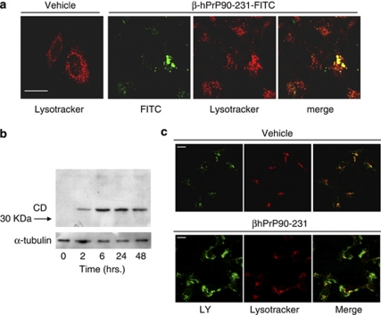
Intracellular β–hPrP90-231 activates endosomal–lysosomal system. (a) β–hPrP90-231-FITC accumulates into endosomal–lysosomal vesicles. Cells were treated with vehicle and β–hPrP90-231FITC (1 μM) for 24 h and counterstained with lysotracker red DND-99, before being observed in live cell confocal microscopy. Cells treated with β–hPrP90-231-FITC show fluorescent round clusters that almost completely colocalize with lysotracker. Vehicle-treated cells were stained with lysotracker red to provide images of lysosomes distribution and size in SH-SY5Y cells. (b) β–hPrP90-231 elicits the expression of active CD. Cells were treated for 2–48 h with hPrP90-231 (1 μM). The amount of active CD (MW 33 kDa) was detected by SDS-PAGE (10%), loading 50 μg of cell proteins/lane. A separate set of samples (10 μg/lane) was probed with the monoclonal anti-α-tubulin to normalize protein content. The expression of 33kDa CD, corresponding to the mature active form, is significantly increased after 2 h, reaches a sustained plateau after 6 h, and lasts up to 48 h of treatment. (c) β–hPrP90-231 treatment causes loss of lysosome membrane impermeability. Cells were treated for 3 days with vehicle (PBS) or β–hPrP90-231 (1 μM). In the last 15 h of treatment, cells were loaded with 100 μg/ml of LY. At 15 min before confocal microscope analysis, cells were loaded with lysotracker red DND-99 (50 nM). Control cells evidenced a vesicular distribution of LY that almost completely overlaps with lysotracker fluorescence, indicating an efficient loading of LY in endosomal–lysosomal vesicles (upper panels). β–hPrP90-231-treated cells exhibited a diffuse LY staining within the cytoplasm outside lysotracker-positive vesicles (lower panel), indicating that LY has been redistributed in the cytosol following the loss of lysosomal membrane impermeability. (Scale bars=10 μm)

