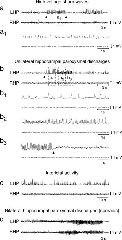
FIG. 2.
Representative electroencephalographic (EEG) tracings of chronic epileptic activity induced by intra-hippocampal kainate in mice. Chronic epileptic activity develops in mice 3 to 7 days after unilateral intra-hippocampal injection (left hippocampus [LHP]) of kainate (200 ng in 50 nl) causing SE. Tracings in (a) (boxed area is enlarged in a1) and (b) (boxed areas are enlarged in b1, b2, b3) depicting high-voltage sharp waves (HVSW) (1–4 mV; 3–8 Hz; average duration, 20 seconds) and unilateral hippocampal paroxysmal discharges (HPDs) that typically start (arrowhead in panel b) with large amplitude sharp waves (1–3 mV; 1–3 Hz; panel b1) followed by a train of spikes of increasing frequency (0.5–1.0 mV; 10–20 Hz, panels b2, b3), and terminating with a deflection in the EEG (arrowhead in panel b3). HVSW and HPDs were inhibited by VX-765 administration (see FIG. 4b–d). Panel (c) depicts interictal activity consisting of isolated spikes or spike trains (1–3 Hz; duration, <20 seconds); panel (d) shows bilateral hippocampal paroxysmal discharges (sporadic events in this model). The events in panels (c) and (d) were not included in the quantification of chronic epileptic activity. RHP = right hippocampus.

