Figure 1.
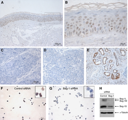
Bag-1 immunohistochemistry of normal human skin. (A) Bag-1 expression in normal human skin. (B) A higher power image of boxed region in A, illustrating predominantly nuclear localisation of Bag-1. Negative controls were: (C) negative control tumour section without primary antibody; corresponding Bag-1 stained tumour showed 80% positive cells (assigned staining intensity scores of nuclear +++, cytoplasmic ++) and (D) rabbit pre-immune serum; corresponding Bag-1 stained tumour showed 80% positive cells (assigned staining intensity scores of nuclear +++, cytoplasmic ++). (E) Strong Bag-1 staining in sweat glands was used as an internal positive control to ensure consistent staining. (F, G and H) Validation of Bag-1 TB3 antibody using small interfering RNA (siRNA) knockdown in the HaCaT keratinocyte cell line. HaCaT cells were transfected with Silencer control siRNA (F) or Bag-1 siRNA (G). (H) Western blot showing reduced expression of Bag-1 protein in HaCaT cells transfected with Bag-1 siRNA compared with Silencer control siRNA. Images A and E were photographed using a × 10 objective (scale bar shown in A), images C, D, F and G were photographed using a × 20 objective (scale bars shown in C and F), and image B using a × 40 objective (scale bar shown).

