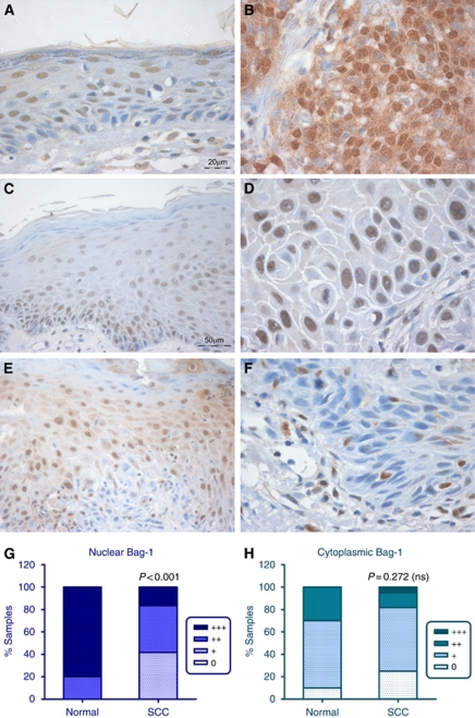
Figure 2.
Expression of Bag-1 in epidermal SCC and adjacent epithelium. (A, C and E) Bag-1 expression in epithelium adjacent to SCC; note the altered pattern of Bag-1 expression in adjacent epithelium, compared with the normal skin expression pattern shown in Figures 1A and B. (B) Moderate to poorly differentiated SCC showing increased cytoplasmic and nuclear Bag-1 expression relative to adjacent epithelium (A). (D) Well-differentiated SCC showing reduced cytoplasmic and increased nuclear Bag-1 expression, compared with adjacent epithelium (C). (F) Well-differentiated SCC showing reduced Bag-1 expression relative to adjacent dysplastic epithelium (E). Images C and E were photographed using a × 20 objective (scale bar shown in C), and images A, B, D and F were photographed using a × 40 objective (scale bar shown in A). (G) Comparison of intensity of Bag-1 nuclear staining in normal epidermis compared with the panel of SCCs. (H) Comparison of Bag-1 cytoplasmic staining in normal epidermis compared with the panel of SCCs. Statistical analysis was performed using a Mann–Whitney test.

