Figure 3.
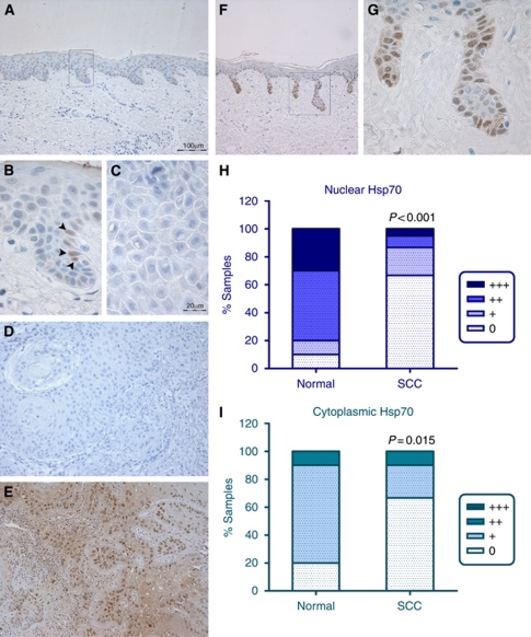
Hsp70 immunohistochemistry of normal human skin, epidermal SCC and adjacent epithelium. (A) Normal epidermal epithelium is predominantly negative for Hsp70 staining, with occasional Hsp70-positive cells, located within the basal layer. A higher power image of the boxed area in A is shown in B, to highlight the presence of occasional Hsp70-positive cells (arrowheads). (C) Negative control (normal goat serum in PBS) tumour section without primary antibody; the corresponding Hsp70 stained tumour showed positive staining in 30% of the cells (assigned staining intensity scores of nuclear ++, cytoplasmic +). (D) Tumour negative for Hsp70 staining. (E) Hsp70-positive staining human epidermal SCC. (F and G) Deep peg-like rete ridges stained strongly positive for Hsp70 in sun-damaged epidermis adjacent to SCC; boxed area in F is shown at higher magnification in G. (H) Intensity of Hsp70 nuclear staining in normal epithelium and in the panel of SCCs. (I) Intensity of Hsp70 cytoplasmic staining in normal epithelium and in the panel of SCCs. Images A, F, D and E were photographed using a × 10 objective (scale bar shown in A). Images B, C and G were photographed using a × 40 objective (scale bar shown in C). Statistical analysis was performed using a Mann–Whitney test. 0=undetectable Hsp70 staining; +=weak; ++=moderate; +++=strong Hsp70 staining.

