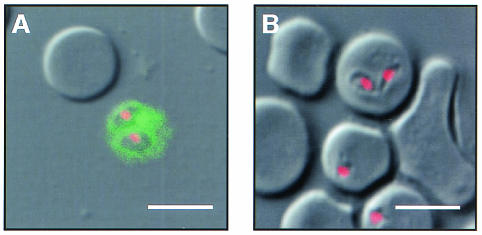
FIG. 3.
Demonstration of heparin binding on the surfaces of free merozoites. B. bovis-infected RBCs were preincubated with heparin-FITC, followed by fixation with acetone-methanol, and were then examined under a confocal laser scanning microscope. The heparin-antigen reaction (green) and nucleus (red) were visualized by staining with FITC and PI, respectively. A diffuse fluorescence reaction was detectable only around free merozoites (A) and not on the infected RBCs (B). Bars, 5 μm.

