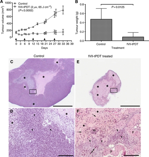
Figure 4.
Factor VII-targeted photodynamic therapy (fVII-tPDT) is effective in the treatment of chemoresistant breast cancer MCF-7/MDR tumours in nude mice. (A and B) After fVII-tPDT treatment (2 μM, 65 J cm–2; arrows indicating the treatment dates), MCF-7/MDR tumour volume (A) and tumour weight (B) from fVII-tPDT-treated mice were significantly smaller than those from control mice. (C–F) Histopathology of MCF-7/MDR tumours from control and fVII-tPDT-treated mice. MCF-7/MDR tumours from control mice (C and D) were large and had multiple variably sized foci of necrosis and cellular debris (*) in contrast to the tumours from fVII-tPDT-treated mice (E and F) that were smaller and composed predominantly of necrotic cellular debris (*). At increased magnification, tumour cells from MCF-7/MDR tumours (D, inset of C) were numerous, easy to identify by their dark blue euchromatic nuclei. In contrast, distinct tumour cells from fVII-tPDT-treated (F, inset of E) mice were markedly fewer in number and in the process of necrosis or apoptosis as evidenced by the diffuse nuclear pkynosis (arrowheads), including the nuclei of large tumour cells (double arrow). Tumour cells were enmeshed in the background of necrotic cellular debris (*) admixed with haemosiderin-laden macrophages (m) and fibroblasts (arrows). Haematoxylin and eosin (HE), scale bars for (C and E)=2000 μm; for (D and F)=200 μm. p=panniculus carnosus muscle.

