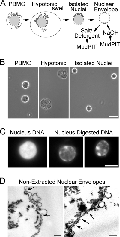
Fig. 1.
Cellular fractionation of PBMCs. A, method schematic. Crude nuclei prepared from hypotonic lysis of PBMCs were cleaned of contaminating cellular structures by gradient centrifugation to float contaminating membranes while pelleting the denser nuclei. Crude NEs were prepared by digesting/extracting nuclear contents from isolated nuclei. Before MudPIT analysis, these were further extracted with 1% β-octyl glucoside, 400 mm NaCl or 0.1 n NaOH to enrich for proteins associated with the insoluble lamin polymer or integral membrane proteins, respectively. B, buffy coats from human blood were separated on Ficoll-Hypaque density gradients to enrich for mononuclear leukocytes (left panel). The cells were swollen hypotonically (middle panel) and Dounce homogenized to release nuclei (right panel), which were then further purified on sucrose gradients. Phase-contrast light microscope images are shown. Scale bar, 10 μm. C, enrichment for NEs by chromatin digestion. DAPI staining for DNA visualizes significant nuclear chromatin content in an isolated PBMC nucleus (left panel) and the loss of most of this material after two rounds of digestion with DNase and RNase, each followed by salt washes (two right panels). A fluorescence microscope image is shown. Scale bar, 5 μm. D, ultrastructure of isolated NEs. Electron micrographs of PBMC NEs show that most membranes in the population are the characteristic double membrane with little contamination from single membrane vesicles. Arrows point to positions of NPCs. In some places, the hypotonic treatment used to swell the NEs while digesting/extracting most of the chromatin resulted in membrane blebbing. These NEs were further extracted with salt and detergent to enrich for proteins associated with the intermediate filament lamin polymer or with an alkaline treatment to enrich for transmembrane proteins prior to analysis by MudPIT. After such treatment, no structure remains that can be readily discerned by EM with the characteristics of NEs. Scale bar, 0.2 μm.

