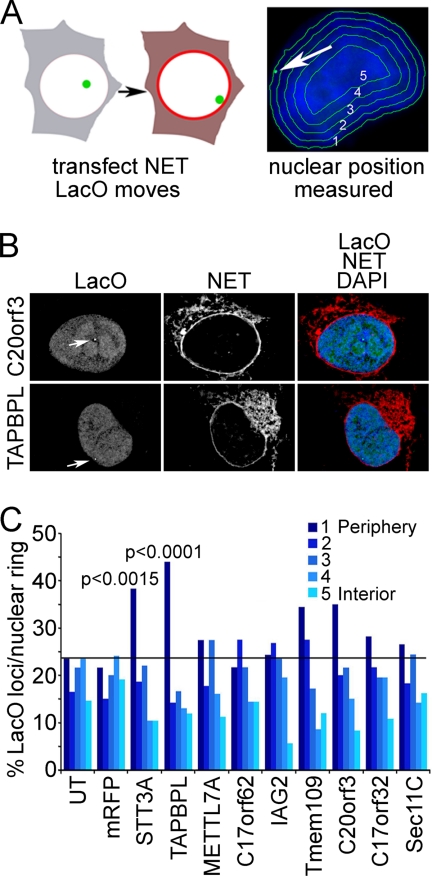
Fig. 7.
Cell-based screen for PBMC NETs that promote recruitment of chromosome loci to nuclear periphery. A, schematic representation of a screen to determine whether overexpression of a particular NET can recruit a specific chromatin locus to the NE. NETs were transfected into cells containing a lacO repeat integration that is typically in the interior. The lacO position was visualized with GFP-lacI (green) and measured using an algorithm that partitions the nucleus based on DAPI staining (blue) into five concentric circles of roughly equal area. B, example of NET-transfected cells. The position of the lacO locus is highlighted by the white arrows. The lacO locus position is unaffected by C20orf3 expression but moves to the periphery with TAPBPL expression. DAPI staining added to the merged image confirms that the movement of the locus is not an artifact of generalized chromatin condensation at the periphery. Deconvolved images are shown. C, the ring containing the locus was recorded in roughly 100 transfected cells. p values were calculated for NETs that increased the locus at the periphery in comparison with untransfected (UT) or mRFP-transfected control cells using a χ2 test.

