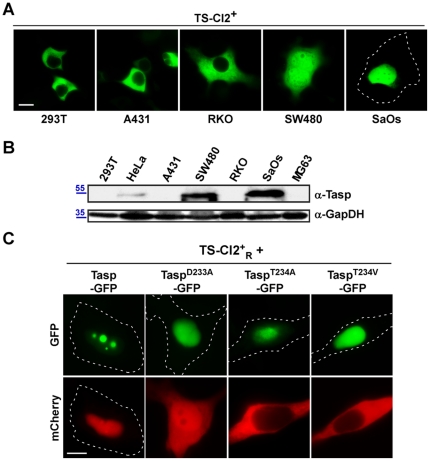
Figure 4. Biosensor-based probing of Taspase1 function in vivo.
A./B. Cleavage of the biosensor correlated with endogenous Taspase1 levels in adherent tumor cell lines. A. Indicated cell lines were transfected with equal amounts of TS-Cl2+ expression plasmid. 24 h later, localization of the biosensor was analyzed in at least 200 fluorescent cells displaying similar fluorescence intensity. Representative examples are shown. Cleavage-induced nuclear translocation differed significantly among tested cell lines. B. Endogenous Taspase1 levels were analysed by immunoblot using α-Tasp and -GAPDH Abs. C. Biosensor-based analysis of the proteolytic activity of Taspase1 variants in HeLa transfectants. Coexpression of TaspT234A- or TaspT234V-GFP fusion did not result in cleavage and nuclear accumulation of TS-Cl2+R. TaspD233A-GFP displayed a reduced enzymatic activity compared to wt Taspase1-GFP. Scale bars, 10 µm. Dashed lines mark nuclear/cytoplasmic cell boundaries obtained from the corresponding phase contrast images.

