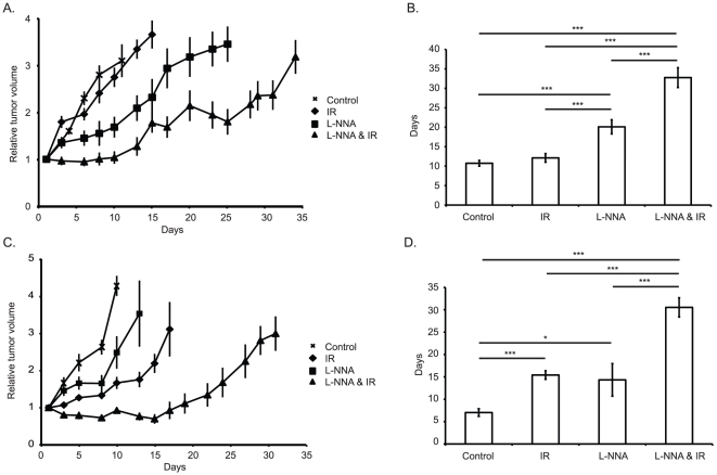
Figure 1. L-NNA and IR reduce the rate of xenograft growth.
Relative volumes of differently treated A431 (A) and FaDu (C) xenografts. Mean tripling time of A431 (B) and FaDu (D) xenografts. Xenografts were created in athymic nu/nu mice by subcutaneous injection of 106 A431 or 5×10⁵ FaDu cells into the hind flank, once tumors were established animals were divided into 4 groups as indicated. L-NNA was provided from day 0 at 0.5 g/L in drinking water, irradiated animals received a single 10 Gy dose targeted at the flank using a 60Co source on day 1. Data presented as mean relative volume ± SEM or mean days to triple volume ± SEM; N = 5, n = 10. * = p<0.05, *** = p<0.01. Horizontal lines and accompanying asterisks indicate statistically significant differences.

