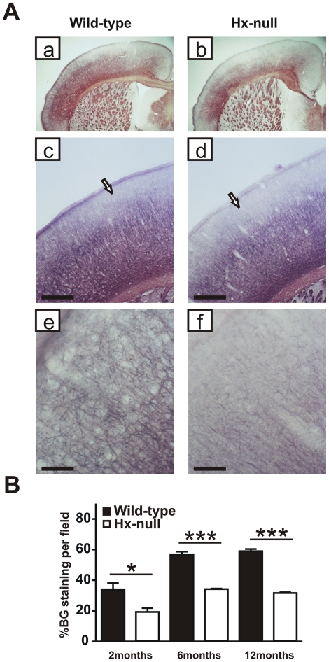
Figure 2. The cortex of Hx−/− mice is hypomyelinated.
A) Coronal sections of a wild-type and a Hx−/− mouse at twelve months of age stained with Black-Gold reaction to detect myelinated fibers. Hx−/− mouse shows reduced myelination in cerebral cortex compared to wild-type (a, b) and the hypomyelination mainly affects the supragranular layers in motor and somatosensory cortex (arrows in c, d). Higher magnification shows that in layer I of Hx−/− mouse the staining is very weak compared to wild-type (e, f). Bar (c, d) = 500 µm; Bar (e, f) = 100 µm. B) Quantification of fiber density in motor cortical area, assessed at 2, 6 and 12 months of age, shows a severe reduction in Hx−/− mice. Data represent mean ± SEM, n = 3 mice for each genotype. * = P<0.05, *** = P<0.001.

