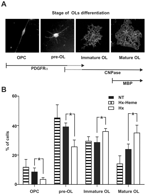
Figure 6. Hx promotes OL differentiation.
OPCs were grown with or without Hx and the differentiation process was analyzed. A) Representative images showing the different developmental stages taken into consideration: stage I, OPCs (bipolar); stage II: pre-OL (primary branched); stage III: immature OL (secondary branched); stage IV: mature OL (secondary branched cells with membranous processes). Cells at stage I and II are PDGFRα positive, CNPase negative, cells at stage III are PDGFRα negative, CNPase positive and cells at stage IV are PDGFRα negative, CNPase and MBP positive. B) Kinetics of OL differentiation. Cells were cultured for 48 h in the absence (NT) or presence of Hx (Hx) or heme-Hx complex (Hx-heme), and the number of cells at each differentiation stage was counted as reported in Materials and Methods. Cells were scored by morphology and immunoreactivity to PDGFα and CNPase as shown in (A). Hx treatment accelerated the differentiation process whereas the heme-Hx complex was ineffective. * = P<0.05. Results shown are representative of three independent experiments.

